Description
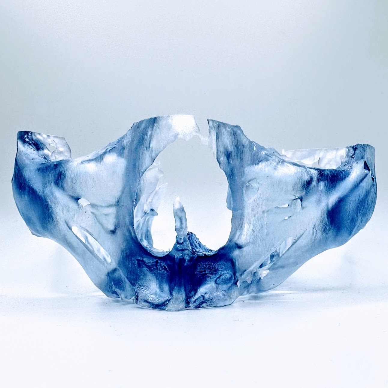
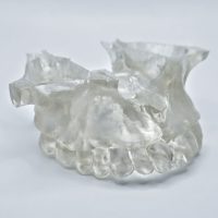
Upper Maxilla Model ONLY – Zygomatic Treatment
This full arch maxilla model includes maxilla, suborbital rim, and zygomatic bone. May include pterygoid bone if visible in CT scan.
Requirements:
☐ CT Scans
Partially/fully dentate:
- CBCT scan of patient with separated occlusion. Use cotton rolls or similar materials as necessary to prop bite open.
- High quality resolution of 0.3 voxel or better
- At least one full arch per scan must be captured. If double arch treatment and the scan volume is small, scan the patient twice with one arch per scan
Fully edentulous:
- CBCT scan of patient wearing relined denture featuring radiographic markers at MIP against opposing
- CBCT scan of the denture only featuring radiographic markers
- High quality resolution of 0.3 voxel or better
- At least one full arch per scan must be captured. If double arch treatment and the scan volume is small, scan the patient twice with one arch per scan
If planning implants:
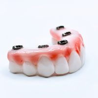
☐ Digital (STL format) or conventional impressions
☐ Digital (STL format) or conventional bite at desired restorative vertical (MIP or corrected centric)
☐ Digital photos of patient (9 photos):
- Full face maximum smile
- Left & right maximum smile
- Full face retracted
- Left & right retracted
- Full face reposed
- Left & right reposed


